Analyst. Perfectionist. Visionary. Organizer.
Pentacam® HR ‒ the Analyst
YOUR BENEFITS
- Premium image quality
- Early detection of ectatic diseases
- Objective keratoconus progression analysis
- Easy fitting of RGP lenses and scleral lenses
INVESTIGATIONS
- Scheimpflug-based tomography
- CSP scan (cornea scleral profile)
SUITABILITY
- Ophthalmologists
- Refractive and cataract surgeons
- Contact lens specialists
- Optometrists
Pentacam® AXL ‒ the Perfectionist
YOUR BENEFITS
- Biometry plus tomography in one device
- Delegable measurement process
- Customized IOL selection and IOL power calculation
- Built-in IOL Calculator
with customized Barrett & Olsen ray tracing formulas
for post-refractive eyes, incl. PCA - Built-in IOL database
INVESTIGATIONS
- Scheimpflug-based tomography
- Optical biometry
SUITABILITY
- Ophthalmologists
- Cataract surgeons
- Refractive surgeons
- Eye care specialist
Pentacam® AXL Wave ‒ the Visionary
YOUR BENEFITS
- Unrivalled pre-op care for your patients
- Customized IOL selection and
- IOL power calculation
- Comprehensive post-op evaluation
- Initial impression of individual
visual performance - Time and space saving
INVESTIGATIONS
- Scheimpflug-based tomography
- Objective refraction
- Wavefront aberrometry of the entire eye
- Optical biometry
- Retroillumination
SUITABILITY
- Ophthalmologists
- Cataract surgeons
- Refractive surgeons
- Eye care specialist
Pentacam® ‒ the Organizer
YOUR BENEFITS
- Anterior segment tomography in 2 seconds
- Scientifically evaluated displays for screening
- Early detection and objective monitoring of ectatic diseases
- Customizable software
INVESTIGATIONS
- Scheimpflug-based tomography
- Total Corneal Refractive Power (TCRP)
- Corneal thickness
SUITABILITY
- Ophthalmologists
- Refractive and cataract surgeons
- Contact lens specialists
- Optometrists
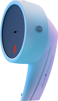
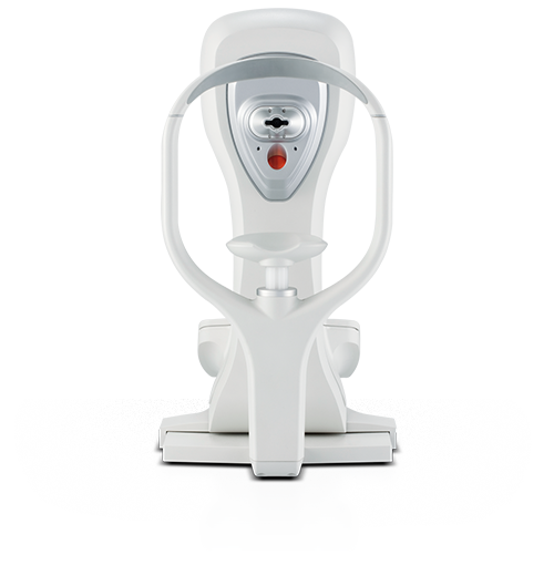
Pentacam® HR
High-resolution Scheimpflug images
of the anterior eye segment
Automatic measurement activation (delegable)
Total Corneal Refractive Power
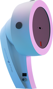
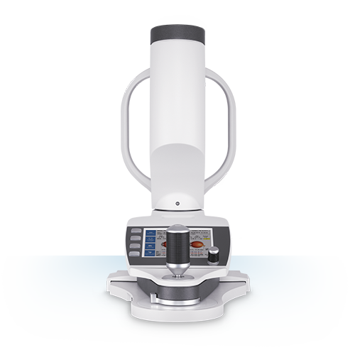
Pentacam® AXL
Anterior segment tomography
and optical biometry
Measurement of axial length
Built-in IOL calculation formulas
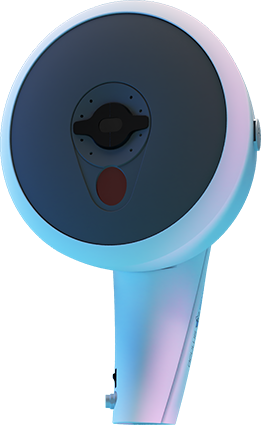
Pentacam® AXL Wave
The next generation
Axial length and refraction
Total wavefront and retroillumination
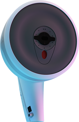
Pentacam®
The Gold Standard for
anterior segment tomography
Comprehensive analysis
Scientifically reliable screenings
FAQs
It stands for the integrated high-resolution camera, based on Scheimpflug technology – for premium image quality!
A Scheimpflug scan produces 50 images of the anterior eye segment or 100 images of the cornea within 2 seconds. It gives you an overall view of the anterior eye segment.
Yes, it measures corneal low and higher order wavefront aberrations.
Yes, every Scheimpflug image can be individually displayed and evaluated by the software. This makes it easier to detect symptoms indicative of Fuch's endothelial dystrophy or other pathologies of the anterior eye segment, for example.
Yes, the IOL Calculator can be added optionally. This software module includes various IOL formulas and more than 350 IOL geometries. Alternatively, links to various external IOL calculators can be set up manually.
No. The Pentacam® AXL has a different hardware. However, some software modules can be added optionally.
FAQs
The Pentacam® AXL uses optical biometry (partial optical coherence interferometry) to measure axial length.
A Scheimpflug scan produces 50 images of the anterior eye segment or 100 images of the cornea within 2 seconds. It gives you an overall view of the anterior eye segment. A combined scan of Scheimpflug tomography and optical biometry (axial length) takes around 2 minutes.
Yes, it measures corneal low and higher order wavefront aberrations.
Yes, every Scheimpflug image can be individually displayed and evaluated by the software. This makes it easier to detect various kinds of corneal or lens opacities, for example.
No. The Pentacam® AXL Wave has different hardware.
FAQs
The measurement process takes around 4 minutes for both eyes. It includes wavefront aberrometry, objective refraction, retroillumination, axial length and Scheimpflug tomography.
The Pentacam® AXL Wave uses Hartmann-Shack technology to measure low and higher order aberrations of the entire eye. It also displays the internal and total corneal wavefront aberrometry results for detailed crystalline lens or IOL assessment.
The implemented retroillumination enables a convenient check of the inclination and centration of IOLs, especially toric IOLs.
Yes, every Scheimpflug image can be individually displayed and evaluated by the software. This makes it easier to detect symptoms indicative of corneal ectasia or changes of the crystalline lens.
Yes. The integrated retroillumination, tomography and wavefront aberrometry functions display most changes in corneal shape, as well as improvements in vision, following refractive or cataract surgery.
FAQs
Scheimpflug-based corneal tomography is the gold standard in anterior eye segment tomography. It is fast, delegable, precise, contact-free and tear-film independent.
A Scheimpflug scan produces 50 images of the anterior eye segment within 2 seconds. It gives you an overall view of the anterior eye segment.
Yes, it measures corneal low and higher order wavefront aberrations.
Yes, every Scheimpflug image can be individually displayed and evaluated by the software. This makes it easier to detect changes of the anterior eye segment when screening for glaucoma, for example.
No. The Pentacam® HR has a different hardware. However, some software modules can be added optionally.
OCULUS Online Show
What Pentacam® type are you?
Pentacam® HR
High-resolution Scheimpflug images
of the anterior eye segment
Automatic measurement activation (delegable)
Total Corneal Refractive Power
Pentacam® HR ‒ the Analyst
YOUR BENEFITS
- Premium image quality
- Early detection of ectatic diseases
- Objective keratoconus progression analysis
- Easy fitting of RGP lenses and scleral lenses
INVESTIGATIONS
- Scheimpflug-based tomography
- CSP scan (cornea scleral profile)
SUITABILITY
- Ophthalmologists
- Refractive and cataract surgeons
- Contact lens specialists
- Optometrists
FAQs
It stands for the integrated high-resolution camera, based on Scheimpflug technology – for premium image quality!
A Scheimpflug scan produces 50 images of the anterior eye segment or 100 images of the cornea within 2 seconds. It gives you an overall view of the anterior eye segment.
Yes, it measures corneal low and higher order wavefront aberrations.
Yes, every Scheimpflug image can be individually displayed and evaluated by the software. This makes it easier to detect symptoms indicative of Fuch's endothelial dystrophy or other pathologies of the anterior eye segment, for example.
Yes, the IOL Calculator can be added optionally. This software module includes various IOL formulas and more than 350 IOL geometries. Alternatively, links to various external IOL calculators can be set up manually.
No. The Pentacam® AXL has a different hardware. However, some software modules can be added optionally.
Pentacam® AXL
Anterior segment tomography
and optical biometry
Measurement of axial length
Built-in IOL calculation formulas
Pentacam® AXL ‒ the Perfectionist
YOUR BENEFITS
- Biometry plus tomography in one device
- Delegable measurement process
- Customized IOL selection and IOL power calculation
- Built-in IOL Calculator
with customized Barrett & Olsen ray tracing formulas
for post-refractive eyes, incl. PCA - Built-in IOL database
INVESTIGATIONS
- Scheimpflug-based tomography
- Optical biometry
SUITABILITY
- Ophthalmologists
- Cataract surgeons
- Refractive surgeons
- Eye care specialist
FAQs
The Pentacam® AXL uses optical biometry (partial optical coherence interferometry) to measure axial length.
A Scheimpflug scan produces 50 images of the anterior eye segment or 100 images of the cornea within 2 seconds. It gives you an overall view of the anterior eye segment. A combined scan of Scheimpflug tomography and optical biometry (axial length) takes around 2 minutes.
Yes, it measures corneal low and higher order wavefront aberrations.
Yes, every Scheimpflug image can be individually displayed and evaluated by the software. This makes it easier to detect various kinds of corneal or lens opacities, for example.
No. The Pentacam® AXL Wave has different hardware.
Pentacam® AXL Wave
The next generation
Axial length and refraction
Total wavefront and retroillumination
Pentacam® AXL Wave ‒ the Visionary
YOUR BENEFITS
- Unrivalled pre-op care for your patients
- Customized IOL selection and
- IOL power calculation
- Comprehensive post-op evaluation
- Initial impression of individual
visual performance - Time and space saving
INVESTIGATIONS
- Scheimpflug-based tomography
- Objective refraction
- Wavefront aberrometry of the entire eye
- Optical biometry
- Retroillumination
SUITABILITY
- Ophthalmologists
- Cataract surgeons
- Refractive surgeons
- Eye care specialist
FAQs
The measurement process takes around 4 minutes for both eyes. It includes wavefront aberrometry, objective refraction, retroillumination, axial length and Scheimpflug tomography.
The Pentacam® AXL Wave uses Hartmann-Shack technology to measure low and higher order aberrations of the entire eye. It also displays the internal and total corneal wavefront aberrometry results for detailed crystalline lens or IOL assessment.
The implemented retroillumination enables a convenient check of the inclination and centration of IOLs, especially toric IOLs.
Yes, every Scheimpflug image can be individually displayed and evaluated by the software. This makes it easier to detect symptoms indicative of corneal ectasia or changes of the crystalline lens.
Yes. The integrated retroillumination, tomography and wavefront aberrometry functions display most changes in corneal shape, as well as improvements in vision, following refractive or cataract surgery.
Pentacam®
The Gold Standard for
anterior segment tomography
Comprehensive analysis
Scientifically reliable screenings
Pentacam® ‒ the Organizer
YOUR BENEFITS
- Anterior segment tomography in 2 seconds
- Scientifically evaluated displays for screening
- Early detection and objective monitoring of ectatic diseases
- Customizable software
INVESTIGATIONS
- Scheimpflug-based tomography
- Total Corneal Refractive Power (TCRP)
- Corneal thickness
SUITABILITY
- Ophthalmologists
- Refractive and cataract surgeons
- Contact lens specialists
- Optometrists
FAQs
Scheimpflug-based corneal tomography is the gold standard in anterior eye segment tomography. It is fast, delegable, precise, contact-free and tear-film independent.
A Scheimpflug scan produces 50 images of the anterior eye segment within 2 seconds. It gives you an overall view of the anterior eye segment.
Yes, it measures corneal low and higher order wavefront aberrations.
Yes, every Scheimpflug image can be individually displayed and evaluated by the software. This makes it easier to detect changes of the anterior eye segment when screening for glaucoma, for example.
No. The Pentacam® HR has a different hardware. However, some software modules can be added optionally.
Program Overview: Enjoy the Show!
Live Online Seminar
Saturday, 3 October 2020, 15-16 h (CEST)
R. Ambrósio Jr, M. Belin

Biomechanics & Tomography
Novel Approaches for Ectasia Screening & Keratoconus Progression Analysis
| Renato Ambrósio Jr, MD | – Artificial Intelligence for Enhancing Scheimpflug Tomography and Biomechanical Assessments |
| Michael Belin, MD | – Clinical Applications of the ABCD Staging and Progression |
Live Roundtable
Wednesday, 28 October 2020, 18-19 h (CEST)
G. Auffarth, T. Kohnen, G. Savini

Anniversary Discount
OCULUS is celebrating its
125 year anniversary this year!
We want you to be part of this
celebration and are giving a 1.25 %
discount* on all OCULUS products.

* The discount only applies to orders made from 1 Oct to 31 Oct 2020. Orders made after 31 Oct 2020 are excluded from this deal. The order needs to be made through an appropriate OCULUS distributor, subsidiary or directly through OCULUS headquarters. The discount only applies, if any contact form on this website was filled out completely and submitted.
Online Seminars
Enjoy expert talks on a huge range of topics:
Diagnostics, refractive and cataract surgery, IOLs, corneal biomechanics and myopia management

By loading the video, you agree to YouTube's privacy policy.
Learn more
Pentacam® AXL Wave – our experiences – worth the investment?
Prof Gerd Auffarth, MD presents his clinical experiences and explains why the Pentacam® AXL Wave is worth the investment.
By loading the video, you agree to YouTube's privacy policy.
Learn more
IOL power calculation with the Pentacam® AXL
Giacomo Savini, MD discusses the Pentacam® AXL with regard to IOL power calculation and current study results.
By loading the video, you agree to YouTube's privacy policy.
Learn more
Pentacam® AXL Wave for refractive cataract surgery
Prof Thomas Kohnen, MD, PhD, FEBO is talking about the role of the new OCULUS Pentacam® AXL Wave for refractive cataract surgery.
By loading the video, you agree to YouTube's privacy policy.
Learn more
The Belin ABCD Keratoconus staging and progression display
Prof Michael Belin, MD explains the science and the clinical applications of Belin ABCD Progression Display.
By loading the video, you agree to YouTube's privacy policy.
Learn more
OCULUS Refractive Camp 1: Tomography meets Biomechanics
Cynthia Roberts and doctors Renato Ambrósio Jr, Paolo and Riccardo Vinciguerra share their knowledge about the importance of corneal tomography and corneal biomechanics for refractive surgery.
By loading the video, you agree to YouTube's privacy policy.
Learn more
OCULUS Refractive Camp 2: Tomography meets Biomechanics
Doctors Ambrósio Jr, Koh, Vinciguerra and Zhou on corneal biomechanics and tomography.
By loading the video, you agree to YouTube's privacy policy.
Learn more
Myopia Management redefined
Philipp Hessler, M. Sc. talks about Myopia Management with the new Myopia Master® from OCULUS.


Act now!
Register for free for our live events,
ask for a quote or contact us for further details.
Get a Quote / Get in Contact
Ask for a quote or just contact us if you need any further information on our products.
* mandatory fields